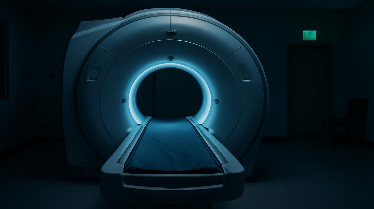
The hum of the machine was softer than you expected, a low mechanical sigh under the bright hospital lights. You lay still on the thin table, arms by your side, trying to ignore the cool air against your skin and the faint smell of disinfectant. A radiology tech smiled above you, adjusting a lead apron, and said something reassuring about how it would all be over in a few minutes. You nodded, but the question was already there, pulsing louder than the scanner’s whirring: Is this safe? Could this scan actually be doing me harm?
The Quiet Fear Behind the Click of the Scanner
A surprising number of us have had that same flicker of doubt. Maybe it flashes across your mind while you’re rolled into an MRI tube or when the CT gantry slides over your chest and the room goes still. Maybe it’s when your child is getting an X-ray after a bad fall from a bike. The fear is simple, blunt, and human: could medical scanners increase the risk of cancer?
To answer that, we have to walk into a strange intersection where physics, biology, and human anxiety all meet. It’s a place full of invisible beams, shielded rooms, and equations about probability rather than destiny. And buried inside all the technical jargon, there is a story about risk, benefit, and how we choose to use some of the most powerful tools in modern medicine.
First, a quiet truth: not all medical scanners are created equal. When we say “scan,” we might mean an X-ray, a CT scan, a PET scan, or an MRI. To your body, those don’t feel the same. Some use ionizing radiation—the kind that can damage DNA. Others use magnetic fields and radio waves that don’t carry enough energy to knock electrons off atoms. To understand your actual risk, we need to sort these out, gently, like separating stones from shells along a shoreline.
What Your Body Actually Feels: Ionizing vs. Non-Ionizing
Imagine standing in a light rain. Each raindrop is like a photon of energy. Most of them are gentle, cool, and harmless; they patter off your jacket and evaporate. But some storms come with hailstones mixed in—hard, fast, and capable of cracking glass. In the world of radiation, ionizing radiation is that hail.
Ionizing radiation has enough energy to knock electrons off atoms and molecules. That’s what makes it both powerful and dangerous. It’s the type used in:
- Conventional X-rays (like chest or dental X-rays)
- CT (computed tomography) scans
- PET (positron emission tomography) scans
- Nuclear medicine scans (bone scans, heart perfusion scans, etc.)
Non-ionizing radiation, by contrast, is like a steady, soft drizzle. It includes:
- MRI (magnetic resonance imaging), which uses strong magnetic fields and radio waves
- Ultrasound, which uses high-frequency sound waves
From a cancer-risk perspective, those last two—MRI and ultrasound—are the quiet good guys. They don’t damage DNA in the way that ionizing radiation can. When people talk about “scanners” giving them cancer, they’re almost always worried about the ionizing kind.
How Medical Radiation Compares: A Pocket-Sized Table
It helps to have something concrete to hold onto. Here’s a compact comparison of typical effective radiation doses and what they roughly mean in terms of additional lifetime cancer risk for an average adult. (These are approximate ranges; actual doses vary by machine, protocol, and body size.)
| Type of Scan | Typical Effective Dose | Rough Extra Lifetime Cancer Risk* |
|---|---|---|
| Chest X-ray | ~0.1 mSv | About 1 in 200,000 |
| Mammogram (both breasts) | ~0.4 mSv | About 1 in 50,000 |
| Head CT | ~2 mSv | About 1 in 10,000–20,000 |
| Abdomen/Pelvis CT | ~8–10 mSv | About 1 in 2,000–2,500 |
| Whole-Body PET/CT | ~20–25 mSv | About 1 in 1,000 |
| MRI | 0 mSv (no ionizing radiation) | No known cancer risk |
| Ultrasound | 0 mSv | No known cancer risk |
*These risk estimates are approximate, based on population averages, and assume the linear no-threshold (LNT) model commonly used in radiation protection.
Can a Scan Actually Start a Cancer? The Biology Behind the Fear
For cancer to begin, something has to go wrong in the DNA of a cell. Ionizing radiation can do this directly, like a tiny invisible bullet cracking the double helix, or indirectly, by creating reactive molecules called free radicals that in turn damage DNA. Most of the time, your body quietly cleans up the mess. Cells repair DNA breaks constantly; it’s part of the ordinary maintenance of being alive.
But sometimes, the repair is imperfect. Sometimes, a misplaced base or a broken chromosome slips through, and if that mutated cell survives and divides, you’ve taken the first microscopic step on a long, uncertain road that could, years or decades later, end in cancer.
This is why ionizing radiation is officially classified as a carcinogen. Not because a single chest X-ray is likely to doom you, but because at high doses—like those experienced by atomic bomb survivors, or workers exposed to heavy radiation—cancer rates clearly rise.
The difficulty lies in the middle ground: what about the small doses from medical imaging? Here, science answers in shades of gray rather than black and white. At very low doses (like a single scan or a few scans), the risk is small—so small that in an individual person, it’s essentially impossible to prove that a particular cancer later came from a particular scan. Instead, researchers look at large populations and use models built from higher-dose data to estimate what might be happening at the lower end.
Most radiation protection frameworks assume a principle called the linear no-threshold model: the idea that any dose, no matter how small, adds a tiny bit of risk, and those little bits add up if we keep exposing ourselves. Is it a perfect model? Probably not. But it’s cautious, and in medicine, cautious is good.
When the Benefit Outweighs the Risk
Picture yourself in an emergency room after a car accident. Your ribs ache when you breathe. There’s a sharp, deep pain in your abdomen that wasn’t there a minute ago. The doctor suspects internal bleeding. A CT scan could reveal a ruptured organ or damaged blood vessel within minutes. If that’s the case, surgery could save your life.
In a moment like that, the long-term theoretical risk of a slightly higher chance of cancer decades later becomes a different creature. You are trading a one-in-a-thousand future risk for a very immediate, very real chance of death today. That’s the central logic of medical imaging: radiation is used because the information it gives us can prevent worse harm.
CT scans are particularly powerful. They can find small tumors, clots in the lungs, tiny brain bleeds, and subtle fractures that plain X-rays miss. PET scans help oncologists stage cancers, see whether treatments are working, and decide whether surgery makes sense. Even humble chest X-rays can spot a collapsed lung or a spreading infection that needs urgent care.
The guiding principle professionals use is sometimes called justification: every test involving radiation should be ordered only when the expected benefit clearly outweighs the risk. For a trauma patient, the calculus is easy. For someone with vague, mild symptoms and a low probability of disease, it becomes much harder—and this is where unnecessary scans creep in.
The Hidden Problem of “Just in Case” Scans
Modern scanners are astonishing. They produce crisp, detailed images in seconds. That convenience can tempt clinicians, health systems, and even patients into a habit of “just in case” imaging: a CT “just to be sure,” a repeat scan “just to check again,” a whole-body screening CT on a perfectly healthy person “for peace of mind.”
But each “just in case” scan sprinkles a little more radiation across your lifetime dose. For an older adult who’s nearing the natural horizon of cancer risk anyway, the added danger is modest. For a child, whose cells are still dividing rapidly and who has many decades ahead, the equation shifts. That’s why pediatric radiology is particularly strict, trimming doses aggressively and avoiding CT when safer alternatives exist.
When scans are used thoughtfully, they save lives. When they become reflex or routine, they can nudge population cancer risks upward in ways that are invisible day-to-day but significant on a national scale. Some studies suggest that a noticeable fraction of future cancers could be linked to medical imaging if scanning practices are not kept in check.
How Doctors Try to Keep You Safe
Radiology departments don’t simply turn machines to full power and hope for the best. Behind the scenes, a quiet engineering and ethical effort is always in motion to squeeze as much information as possible out of the smallest possible dose.
One of the central ideas here is captured in a small acronym with big implications: ALARA, which stands for “As Low As Reasonably Achievable.” It doesn’t mean “as low as imaginable,” because then you might not get a useful picture at all. Instead, it represents a balance: use the minimum radiation that still gives you the diagnostic clarity you need.
This shows up in many small, practical ways:
- Tailored protocols: Children, lighter adults, and specific body parts all have customized settings that reduce dose.
- Modern detectors and software: Newer scanners often need less radiation to create clear images, thanks to better technology and noise-reduction algorithms.
- Shielding and positioning: Technologists use lead aprons or collars when appropriate and carefully position the beam to avoid unnecessary exposure to nearby organs.
- Scan region limits: Only the area of concern is imaged, not the entire body by default.
- Tracking and guidelines: Many hospitals monitor how often and how intensely scans are used, comparing practices to professional guidelines that discourage overuse.
There is also a growing cultural shift within medicine itself. Radiologists and referring doctors are increasingly encouraged to ask, every time: Will this scan change what we do next? If the answer is “no,” that’s a red flag. The best scan is often the one you never have to get at all.
Practical Ways to Protect Yourself Without Avoiding Needed Scans
Knowing that scans carry some risk can make you want to avoid them entirely. But that instinct, while understandable, can be dangerous. Untreated appendicitis, a missed stroke, or a hidden tumor can shorten or end life far more reliably than a theoretical cancer risk decades away.
The goal isn’t to say “no” to imaging; it’s to say “yes” wisely. There are ways you can be an active participant in that process, without needing a physics degree or a medical textbook.
Questions Worth Asking Before You Step into the Scanner
Before agreeing to a scan that uses ionizing radiation—especially a CT, PET, or nuclear medicine study—you can respectfully ask your clinician a few focused questions:
- “How will this scan change what we do?”
If the scan won’t alter diagnosis or treatment, its justification is weak. - “Is there a safer alternative?”
Sometimes MRI or ultrasound can answer the same question with no radiation. - “Have I had similar scans recently?”
In some cases, previous imaging can be reviewed rather than repeated. - “Is this the lowest-dose option for my situation?”
This invites your care team to think about dose-optimized protocols.
Most clinicians appreciate thoughtful questions. You’re not second-guessing their expertise; you’re partnering in the decision. If an emergency leaves no time for discussion, know that in those moments, the immediate risk often vastly outweighs the long-term one. Saving a life now comes first.
It can also help to keep your own quiet record of major scans—especially CT and PET. Even a simple note in your phone listing the type and date is valuable. Over a lifetime, this gives you a clearer sense of your exposure history and can inform conversations with future doctors.
Living with the Tiny Risk: Perspective in an Imperfect World
So, could medical scanners increase the risk of cancer? Yes, in principle and in practice, they can—if they use ionizing radiation. The risk from a single scan is usually very small, often dwarfed by the risk of leaving a serious condition undiagnosed. But across millions of people and billions of images, those tiny risks accumulate into something we can no longer ignore.
At the same time, these same machines have transformed medicine. They reveal strokes in time to dissolve clots, spot aggressive cancers while they are still curable, and guide surgeons with millimeter precision. They reduce uncertainty in ways that spare people from needless operations, or from going home with a lethal problem still festering inside.
Living with medical imaging, then, is a bit like living near a river. The water can nourish, connect, and sustain, but in certain conditions it can flood. The answer is not to abandon the river, but to respect it: build levees, watch the weather, and use it wisely. In hospitals and clinics, that wisdom looks like thoughtful ordering, optimized doses, and open conversations about risk and benefit.
The next time you find yourself on that narrow table, with the machine humming around you and your thoughts racing ahead, remember this: you are not passive in this story. You can ask questions. You can weigh options. And you can hold two truths at once—that this scan carries a tiny shadow of risk, and that it may also be the very thing that keeps you well enough to worry about the future in the first place.
Frequently Asked Questions
Do MRI scans increase the risk of cancer?
No. MRI uses strong magnetic fields and radio waves, not ionizing radiation. There is no credible evidence that MRI increases the risk of cancer. Some people may experience discomfort from noise, claustrophobia, or contrast agents, but cancer is not a known risk.
Are CT scans dangerous?
CT scans use higher doses of ionizing radiation than plain X-rays, so they do carry a small increase in lifetime cancer risk. For most people, the benefit of a medically justified CT—such as diagnosing a serious problem—far outweighs that risk. Problems arise mainly when CT is overused or repeated unnecessarily.
How many scans are “too many”?
There is no single magic number, because the risk depends on dose, type of scan, age, and individual sensitivity. A few CT scans in a lifetime for well-justified reasons are unlikely to cause harm. Large numbers of high-dose scans, especially starting in childhood, can noticeably increase risk. Tracking your imaging history and avoiding unnecessary repeats is a sensible approach.
Should I worry about my child getting a CT scan?
Children are more sensitive to radiation and have more years ahead in which a radiation-related cancer could develop. That’s why pediatric imaging uses special, lower-dose protocols. If a CT is recommended for a child, it’s usually because the information is considered crucial. You can ask whether an MRI or ultrasound could be used instead, but if CT is clearly the best option, the immediate benefit typically outweighs the future risk.
Is a mammogram likely to cause breast cancer?
Mammograms do involve a small amount of ionizing radiation to sensitive tissue, but the dose is low. For most women in recommended age ranges, the benefit of early cancer detection from regular mammography screening far exceeds the slight increase in radiation-related risk.
Do dental X-rays add much to my cancer risk?
Modern dental X-rays use very low doses, especially when digital sensors and proper shielding are used. Occasional dental X-rays contribute only a tiny fraction to your overall lifetime radiation exposure. However, taking them more often than clinically necessary is not helpful, so it’s reasonable to follow evidence-based intervals rather than automatic yearly images.
Can I refuse a scan if I’m worried about radiation?
You always have the right to refuse any test. The key is to understand what you might be giving up. Before deciding, ask your doctor what the scan is meant to find, what could happen if you skip it, and whether a non-radiation alternative exists. A well-informed decision, made together with your care team, is better than a reflexive yes or no.