The first thing Elena noticed was the color green vanishing from her world. Not all at once, not dramatically, just a slow fading—like someone was turning down the saturation on life. The leaves outside her kitchen window dulled, streetlights at night bled into starbursts, and reading a menu became a negotiation with the blur at the center of her vision. Her doctor told her the problem was in the front window of her eye: the cornea. A small scar, he said. A tiny cloud in the clear dome that bends light into focus. Tiny, maybe, but to Elena it felt like a shadow cast over everything.
For years, stories like hers had one main solution: corneal transplant surgery. A long wait for a donor, sutures, risk of rejection, months of recovery. But on a cold morning in a research clinic across the city, another patient lay under a microscope, eyes numb but awake, watching a light shimmer and sharpen as a surgeon slid a nearly invisible gel into a tiny space inside the eye. No big incision. No donor. No stitches. Just a few minutes—and a chance to see clearly again.
When the Eye’s Window Turns Cloudy
The cornea seems almost too simple to hold so much power. It’s just a clear dome of tissue at the front of the eye, thinner than a credit card, curved like a watch glass, and almost entirely transparent. Yet it does most of the bending of light that lets you focus on a bird in the distance or the letters on this screen. When it’s smooth and clear, you don’t think about it at all. You’re not supposed to. Nature designed it to be invisible.
But life has a way of leaving fingerprints on that clarity. A stray branch catches an eye on a hike. A piece of metal from a factory floor flies just a little too far. An infection leaves a milky scar. A genetic condition slowly warps the cornea into a cone shape, like a mountain rising in the wrong place—this is keratoconus. Sometimes even surgery, meant to fix vision, alters the cornea’s shape more than expected.
The result is the same: light no longer glides cleanly through the front window. Instead, it scatters. Straight lines break. Halos appear around headlights. Faces blur just enough to feel more like memories than encounters. Glasses and contact lenses can help for a while, but they’re like trying to smooth a rippled lake with your fingertips. The distortion lives in the tissue itself, in the very architecture of the cornea.
For patients whose corneas are badly scarred or misshapen, doctors have long turned to transplants—carefully replacing the damaged tissue with donated cornea from someone who has died. It’s an astonishing gift. It’s also complicated. Access depends on donor availability, surgical skill, and a health system that can sustain delicate operations. In some parts of the world, people wait years. Elsewhere, the surgeries simply don’t exist.
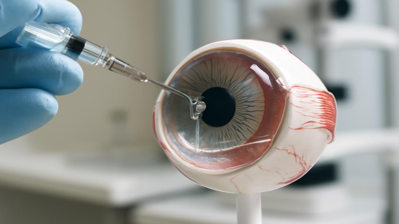
A Quiet Revolution in a Clear Drop
Inside a bright lab with humming refrigerators and sterile glassware, the solution that might change all this doesn’t look like much. It’s a gel—clear, soft, almost ghostlike in a syringe. Hold it up to the light and you see almost nothing at all. But at the microscopic level, it’s a scaffold, a structure that mimics the natural matrix of the cornea, capable of settling into damaged spaces and holding shape like cartilage in a joint or silicone sealant filling a crack in glass.
It isn’t magic. It’s chemistry and biology listening closely to anatomy. The gel is often built around molecules similar to those already found in the cornea: collagen-like structures, or specially designed polymers that are biocompatible and safe. They can be tuned to be just soft enough to match the surrounding tissue, yet stable enough to last. Some versions slowly integrate with the body, inviting the patient’s own cells to move in and remodel. Others act as long-term placeholders—optical prostheses that simply sit there and bend light correctly.
The procedure that uses this gel feels surprisingly understated compared to the drama of cutting and stitching a transplant. Under a microscope, the surgeon makes a small incision at the edge of the cornea—often just a few millimeters long. Through that tiny doorway, fine tools create a pocket within the layers of the cornea, or a shallow space where scar tissue once sat. Then, almost delicately, the clear gel is injected inside.
You can imagine it like slipping a transparent contact lens inside the cornea instead of on top of it. It fills the hollows or pushes aside distortion, rounding out what should be smooth and regular again. As the surgeon shapes it with tiny movements and gentle pressure, the curve of the cornea shifts—subtly, precisely. Light that moments before scattered in confusion now finds a cleaner path toward the retina.
All this happens under local anesthesia. Patients might hear the whir of the microscope or feel a sense of pressure, but not pain. There’s no need to remove the entire front window of the eye, no long, looping stitches, and in many cases, no donor tissue at all.
Stories of Sight, Rewritten in Real Time
In a small exam room, weeks after her procedure, Elena sat with her chin on the rest of a slit lamp—the bright microscope ophthalmologists use to peer into the landscape of the eye. Outside, winter sun reflected off snow in sharp, glittering shards she could actually see, not just sense. The eye doctor flipped through her chart, then through lenses in a handheld frame in front of her face.
“Read the smallest line you can,” he said.
Elena squinted, then relaxed, surprised by how clearly the letters formed. “D… F… Z… N… L…” She laughed halfway through. “I haven’t read that far down since college.”
Her cornea still bore the history of its injury under magnification—a faint trace here, a remodeled layer there—but to her, the world had returned to focus. The gel, carefully injected, had smoothed the optical surface, neutralizing the worst of the distortion. More importantly, she hadn’t spent months in fear of rejection or graft failure. Her recovery had been measured in days and weeks, not seasons.
Elena’s experience is still new and, in many countries, part of clinical trials rather than everyday practice. But her story echoes those of patients with keratoconus whose bulging corneas were reshaped from the inside, and people with small, central scars whose cloudy spots were gently lifted and replaced with clarity. For many, the shift is subtle—a crisper edge on a tree branch, a clearer margin around the moon. For others, it’s the difference between counting fingers and reading street signs.
Doctors who once had to say, “We’ll wait until it’s bad enough for transplant,” are beginning to have a different kind of conversation: one about minimally invasive repair, about preserving a patient’s own cornea for as long as possible, about options that bridge the wide gap between glasses and major surgery.
The New Shape of Treatment Choices
For patients and clinicians alike, this emerging approach changes the decision-making landscape. It adds one more piece to a toolkit that already includes rigid contact lenses, corneal cross-linking to strengthen weakened tissue, laser reshaping in some cases, and, as a last resort, transplant. Now, much earlier in that journey, there can be a question: “Could a gel-based procedure restore enough clarity for this eye?”
In many clinics, conversations begin with comparisons—not just of vision outcomes, but of time, risk, and access. The table below offers a simplified glimpse of how this new technique stacks up against traditional corneal transplant surgery.
| Aspect | Clear Gel Procedure* | Corneal Transplant |
|---|---|---|
| Surgical approach | Small incision, minimal tissue removal | Larger incision, donor tissue replacement |
| Anesthesia | Usually local | Local or general |
| Recovery time | Days to weeks for functional vision | Months for stable vision |
| Donor tissue needed | Typically no | Always yes |
| Risk of rejection | Low (no foreign living tissue) | Present (immune rejection possible) |
| Future options | Transplant still possible if needed later | Revision or repeat graft more complex |
| Stage of disease | Often suitable earlier in disease | Usually for advanced damage |
*Specific techniques and availability vary by country and clinical trial status.
This doesn’t mean the clear gel approach replaces transplants outright. There are still eyes too severely damaged, corneas too thin or irregular, cases where only a full replacement will restore useful sight. But as more patients are treated with minimally invasive methods, the idea of waiting for vision to collapse before acting begins to feel outdated.
The Hidden Engineering of a Gentle Procedure
Behind the apparent simplicity of “injecting a gel” lies a deep collaboration between physics, biology, and patience. The cornea must not only be clear—it must have the right curvature. Think of it like the front lens of a camera. If its surface is even slightly warped, the image blurs. Surgeons planning a gel-based procedure study detailed maps of the patient’s cornea: topography scans that look like colorful weather charts, thickness measurements, and high-resolution cross-sectional images.
With those maps in hand, they plan where to create internal pockets or remove tiny amounts of scar tissue. The gel has to be placed at the right depth to influence the cornea’s shape without compromising its strength. Too close to the surface, and the cornea could become unstable. Too deep, and the optical impact fades. There’s a delicate sweet spot—thick enough to matter, thin enough to stay safe.
The gel itself is engineered to behave more like tissue than like a rigid implant. Some formulations are “shear-thinning,” meaning they flow easily through the thin injection needle but then firm up once in place. Others respond to light, solidifying when exposed to a specific wavelength during surgery. Almost all are designed to be friendly to cells, not triggering aggressive inflammation or rejection.
Surgeons often describe a kind of instant feedback as they work. Under the microscope, with careful illumination, they watch how the cornea’s curve shifts in real time. Instruments can measure changes in focus during the procedure, guiding how much gel to add or how to shape it. It’s like sculpting glass that’s somehow both solid and alive.
Once the eye heals, many patients forget about the gel altogether. They simply live with the new clarity. The body seals the small incision. The tear film glides over the surface. Light slides in, more or less the way nature once intended, before accident or illness intervened.
What It Feels Like to Come Back Into Focus
People often expect a sudden, cinematic moment when the bandages come off, colors bloom, and a choir sings. Reality, as usual, is quieter and more personal. Patients report a mix of tiny revelations: the crispness of text on a phone, the edge of a shadow on a sidewalk, the ability to see their own eyelashes in a mirror without squinting into distortion.
For some, the biggest change is at night. Headlights that once exploded into blinding, starry bursts shrink into more manageable orbs. Street signs no longer smear into fog as they approach. The glowing halo around the moon pulls back, revealing its real shape again.
There’s also the psychological layer: the easing of that low-level anxiety that comes with fragile vision. The constant mental math of “Can I drive?” or “Will I be able to see the slides in that meeting?” begins to relax. Patients who postponed hobbies—birdwatching, painting, woodworking—find themselves drawn back to old loves, no longer held back by uncertainty.
It isn’t perfect for everyone. Some still need glasses or contact lenses for the sharpest vision. Others have modest rather than dramatic improvements. But even incremental changes can feel enormous when they shift daily life from cautious to confident.
Global Echoes: A Technology With World-Spanning Potential
If you step outside the clinic and zoom out to a global view, the potential of restoring sight without major surgery becomes even more striking. In many regions, corneal blindness is common but transplant surgery is rare. Donor tissue is scarce, surgical infrastructure limited, and specialized training hard to come by. The result is a quiet, widespread loss of sight that could, in theory, be prevented or improved.
Gel-based, minimally invasive approaches offer a tantalizing possibility: treatments that are less dependent on donor tissue and elaborate operating rooms. A small incision, a syringe, a specialized gel, and a trained surgeon or eye specialist—if that package can be made affordable and resilient, it could reshape not only individual corneas but entire public health strategies.
Of course, that leap from promising science to widespread access is not automatic. It takes rigorous clinical trials, regulatory approvals, training programs, and careful cost analysis. It requires making sure the materials used are stable in different climates, that follow-up care is manageable, and that patients are informed partners in their treatment. But the direction of travel is clear: away from an all-or-nothing world of “glasses or transplant” toward a spectrum of intermediate, less invasive options.
In the background, another quiet benefit emerges: by reducing dependence on donor tissue for some cases, this technology could free up more donated corneas for those who truly need full transplants. A gel that fills a scar in one eye might indirectly help another patient halfway across the world receive the donor tissue they require.
The Future Glimmering at the Edge of Vision
Look ahead a decade or two, and it isn’t hard to imagine a world where “corneal gel repair” becomes a standard phrase in eye clinics, as familiar as cataract surgery is today. Different formulations might be tailored to different conditions—one type for early keratoconus, another for post-injury scars, a third for subtle irregularities after previous surgeries.
Researchers are already exploring gels that don’t just reshape but also heal: materials infused with growth factors, or designed to release medications slowly over time, helping the cornea regenerate healthier tissue. Some experimental systems aim to work in partnership with light-based treatments, strengthening the cornea at the same time as they smooth it.
Behind all this technical ambition lies something very human: the simple desire to see our lives clearly. To read a child’s expression across a room. To notice the fine details of a leaf after rain. To walk down a street at dusk and trust your own steps. When scientists talk about bioengineered polymers and refractive indices, they’re really talking about those moments.
Elena doesn’t use the word “biomaterial” to describe the gel inside her eye. She just says, “I feel like I’ve been given my window back.” When she stands at her kitchen sink now, looking out at the tree that first told her something was wrong, the green is no longer washed-out, the branches no longer double. The world feels like it has snapped into focus—not with drama, but with a quiet, enduring relief.
Restoring sight without major surgery sounds, at first, like the stuff of science fiction. Yet, drop by clear drop, pocket by tiny pocket, it is becoming something else: an ordinary miracle of modern medicine. Not a grand, headline-grabbing cure, but a gentle reshaping—a way to polish the windows of the eye from the inside, letting the light in again.
FAQ
Is the clear gel procedure available everywhere?
No. In many places, this approach is still part of clinical trials or limited to specialized centers. Availability depends on local regulations, training, and approval of specific materials. An eye specialist can tell you what options currently exist in your region.
Who might be a candidate for this kind of treatment?
Potential candidates often include people with corneal scars, early to moderate keratoconus, or irregular corneas after previous surgery. Suitability depends on corneal thickness, shape, and overall eye health. A detailed exam and imaging are essential.
Does the gel stay in the eye permanently?
It depends on the formulation. Some gels are designed to be long-lasting and remain in place for many years. Others gradually integrate with or are replaced by the body’s own tissue. Your surgeon will explain how the specific material used in your case behaves over time.
Will I still need glasses or contact lenses afterward?
Many patients see significant improvement, but some still need glasses or contacts for the sharpest vision, especially for fine print or night driving. The goal is often to reduce distortion and dependence on strong corrective lenses, not always to eliminate them completely.
Is this safer than a corneal transplant?
“Safer” depends on the individual situation. The clear gel procedure is less invasive and doesn’t use donor tissue, so certain risks—like immune rejection—are lower. However, it also may not be suitable for very advanced disease, where transplant remains the best option. Risk and benefit must be weighed case by case.
How long does recovery usually take?
Most people experience useful vision within days to a few weeks, though the eye can continue to fine-tune and heal over several months. Compared to traditional transplants, the overall recovery period is generally shorter.
Can I still have a corneal transplant later if I need one?
In many cases, yes. One advantage of this minimally invasive approach is that it typically preserves the option of full corneal transplant in the future if the disease progresses or if results are not sufficient. Your surgeon will discuss how the procedure might affect future choices.